A deep dive into the brain
Researchers from ETH Zurich and University of Zurich have developed a new microscopy technique that lights up the brain with high resolution imagery. This allows neuroscientists to study brain functions and ailments more closely and non-invasively.
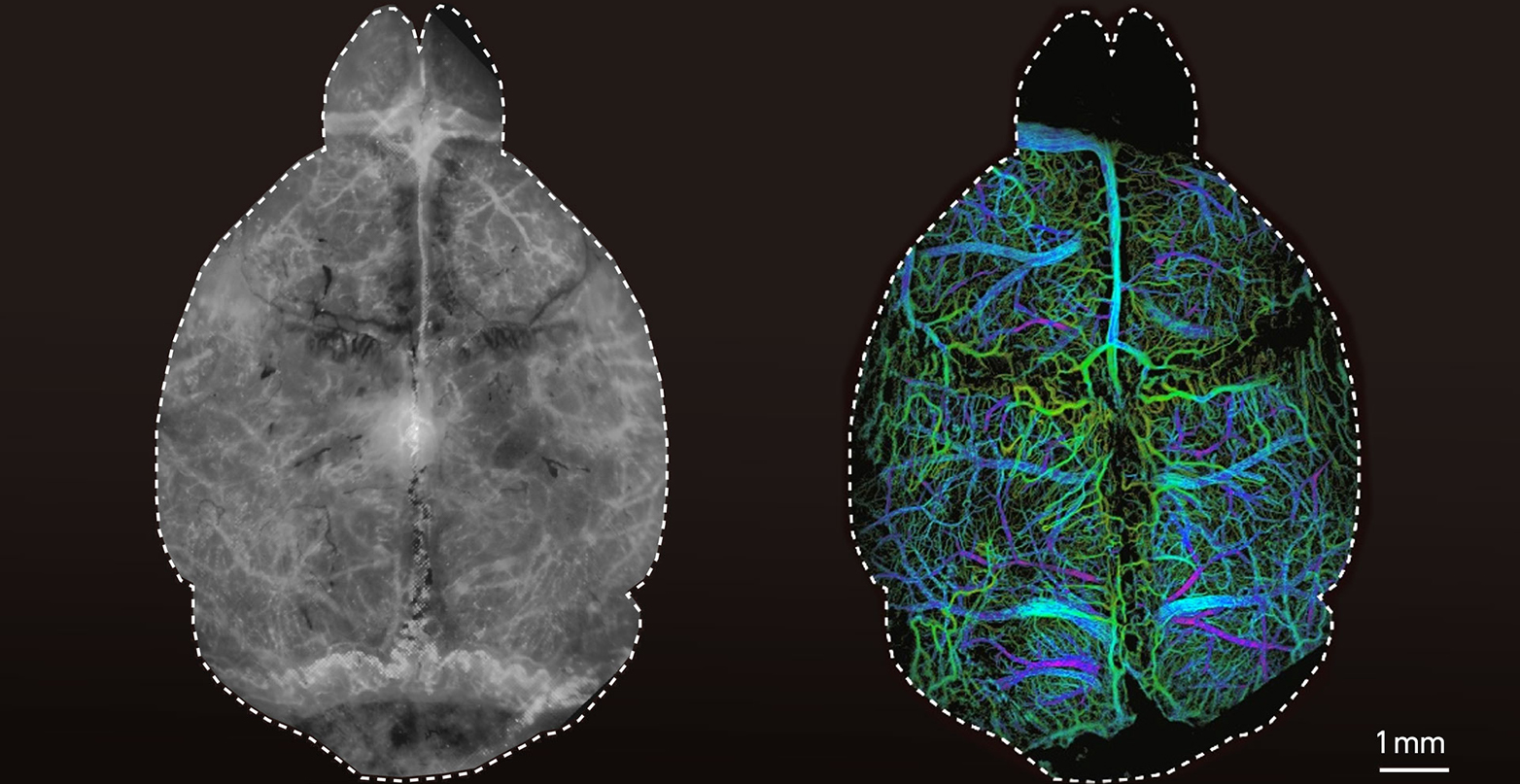
The way the human brain works remains, to a great extent, a topic of controversy. One reason is our limited ability to study neuronal processes at the level of single cells and capillaries across the entire living brain without employing highly invasive surgical methods. This limitation is now on the brink of change.
Researchers led by Daniel Razansky, Professor of Biomedical Imaging at ETH Zurich and the University of Zurich, have developed a fluorescence microscopy technique that facilitates high-resolution images of microcirculation without the need to open the skull or scalp. The technique has been named “diffuse optical localization imaging”, or DOLI in short.
For Razansky, this brings us closer to achieving a long-standing goal in neuroscience: “Visualising biological processes deep in the intact living brain is crucial for understanding both its cognitive functions and neurodegenerative diseases such as Alzheimer’s and Parkinson’s,” he says.