Better cancer diagnosis thanks to digital 3D images
How to bring a diagnostic process that has endured for 100 years into the digital age? Two researchers from ETH Zurich and the University of Zurich are developing a robotic platform that enables a more accurate diagnosis of cancer cells by rapidly quantifying tissue samples in their entirety.
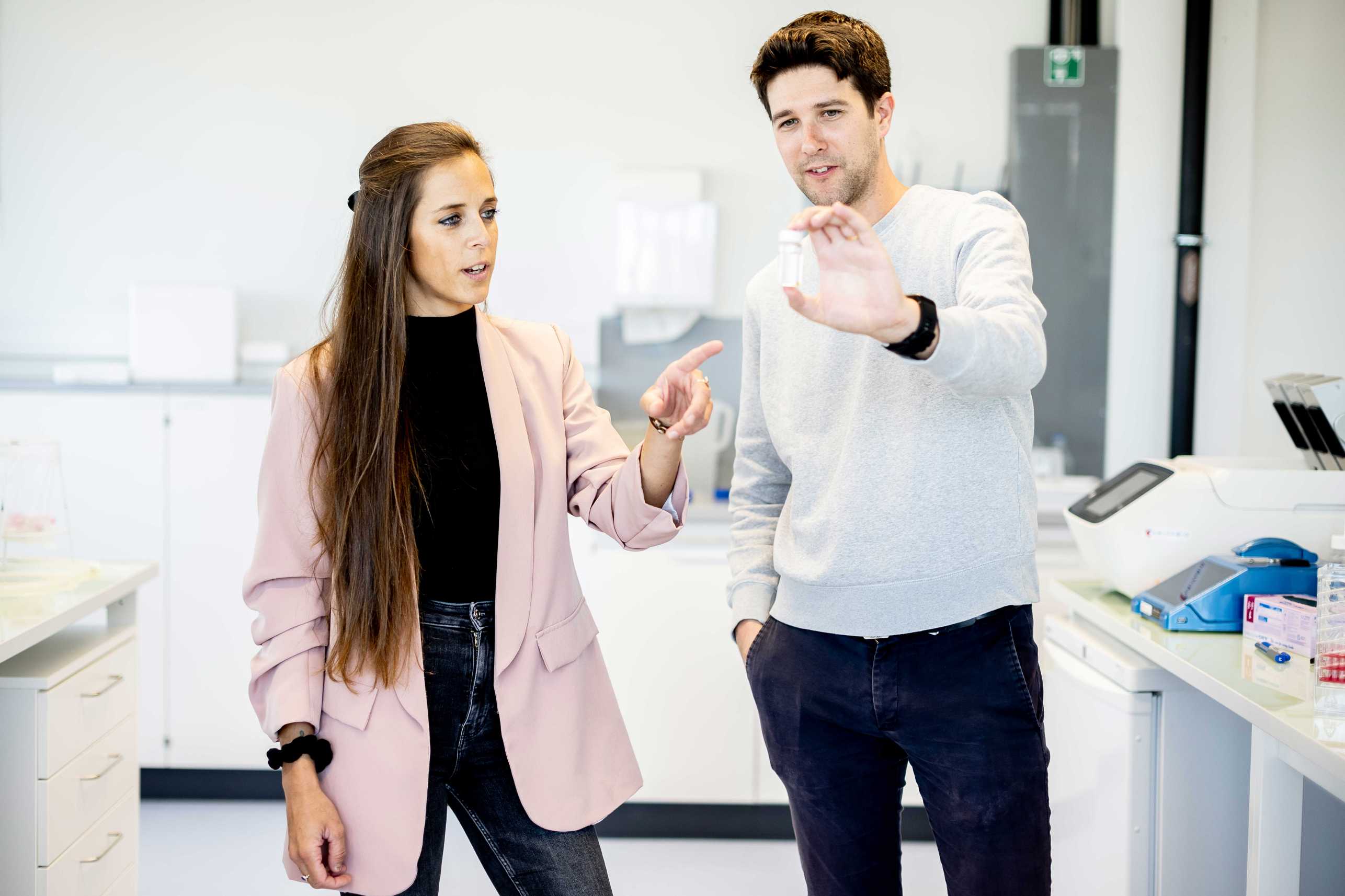
It all started with an innocuous question at the start of Francesca Catto’s doctoral thesis: wouldn’t it be nice if tissue samples could be coloured and digitally displayed as a 3D image? For over 100 years, histology, a branch of pathology that deals with tissue changes, has been using an analogue method that involves cutting tissue samples into micrometre-thin slices (about seven times thinner than a human hair) and examining them for pathological mutations under the microscope. This technique results in one in six people being misdiagnosed and cancer cells going undetected.
Catto, who did her thesis in neuroscience at the University of Zurich under the supervision of Professor Adriano Aguzzi, describes the early days as difficult: “We started out trying different approaches, but nothing worked. It was a nightmare. Fortunately, we entered into a collaboration with the groups of Professor Mirko Meboldt and Professor Alexander Mathys that resulted in the emergence of a successful approach. That’s how Robert Axelrod, who has a doctorate in the field of processing technologies from ETH Zurich, joined the project.”