Visualising multiple sclerosis with a new MRI procedure
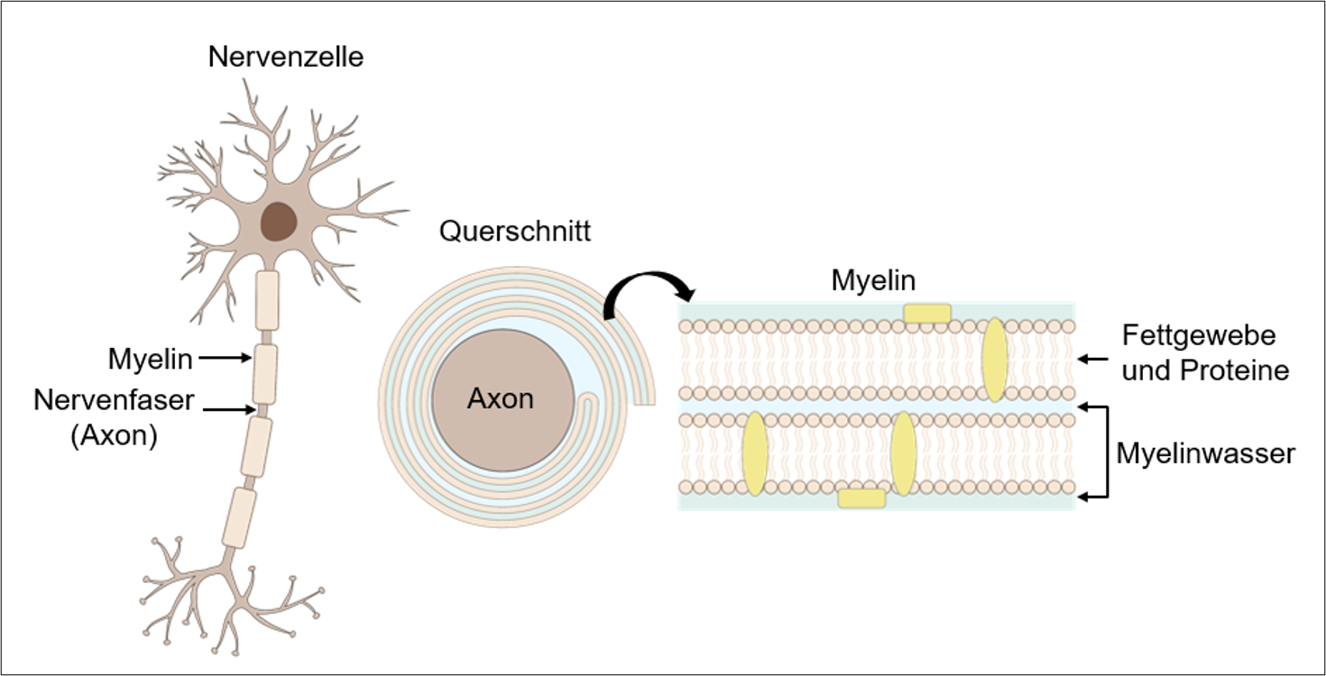
The loss of myelin sheaths in the brain is a hallmark of multiple sclerosis. Researchers at ETH Zurich have now developed an MRI method that maps the condition of this nerve insulation layer more accurately than before.
In brief
- ETH Zurich researchers have developed a new method of magnetic resonance imaging (MRI) for the early detection and better monitoring of multiple sclerosis (MS).
- The method maps the myelin sheaths in the brain more precisely than was previously possible. The loss of myelin sheaths is a hallmark of MS.
- The new MRI method with its special head scanner could also be used by researchers to better visualise other solid tissue types such as connective tissue, tendons and ligaments.